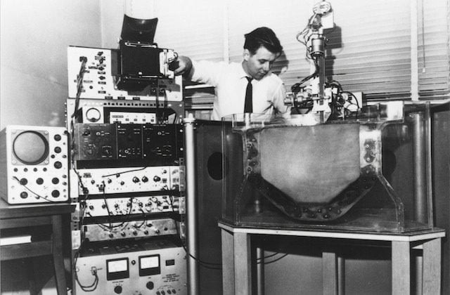
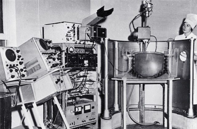
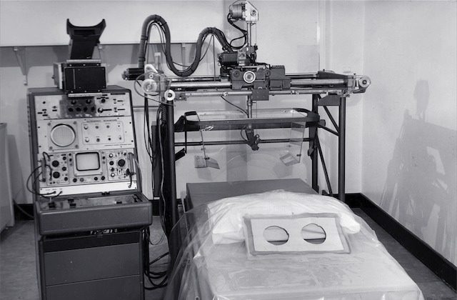
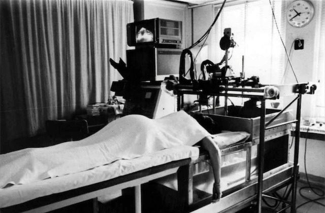
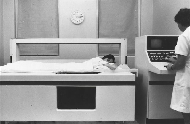
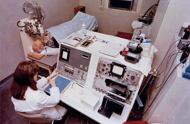
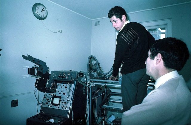
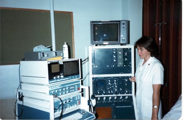
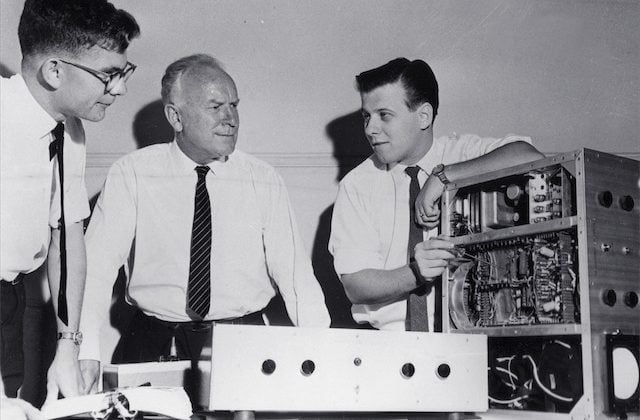
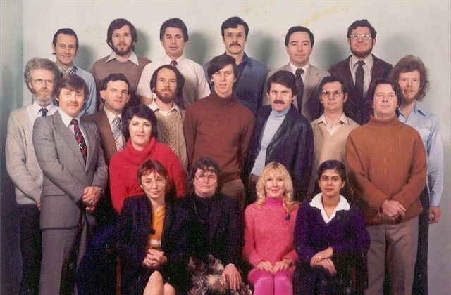
The story of Australian medical ultrasound began 50 years ago with the establishment in 1959 of an Ultrasound Research Section within the Commonwealth Acoustic Laboratories. Within a few years the group was recognised as one of the leading ultrasound research groups in the world.
In 1975 the group became a separate entity and its name was changed to the Ultrasonics Institute (or UI). The Institute contributed vigorously to the development of the technology and clinical applications of diagnostic ultrasound until 1997.
| 1961 | Design and construction of weakly focussing broadband transducers (George Kossoff, Ian Shepherd & Reg Allen) |
| 1962 | First images of late pregnancy at Royal Hospital for Women with Mk I Abdominal Echoscope (David Robinson, George Kossoff, George Radovanovich, Dr William Garrett) |
| 1966 | First images of breast at Royal North Shore Hospital (Jack Jellins, George Kossoff, Brian Hill, Prof. Tom Reeve) |
| 1967 | First images of eye at Royal Prince Alfred Hospital (Michael Dadd, David Robinson, George Kossoff, Dr Bill Hughes) |
| 1967 | Mk II Abdominal Echoscope (David Robinson, George Kossoff, George Radovanovich, Dr William Garrett) |
| 1968 | Publication of comprehensive article on ultrasound image artifacts (David Robinson) |
| 1969 | Development of grey scale imaging, leading to significantly improved images (George Kossoff, David Carpenter, Michael Dadd, Jack Jellins, Kaye Griffiths, Margaret Tabrett) |
| 1972 | National Health Act amended to authorise Ultrasonics Research Section to provide “research and advisory services” to the Department and Government |
| 1973 | First images of the brains of infants (George Kossoff, Margaret Tabrett, Kaye Griffiths, Dr William Garrett, Dr Peter Warren) |
| 1973 | Mini-computer installed to acquire and process ultrasound data (David Robinson, Bruce Williams) |
| 1974 | Curved Annular Phased Array installed on the Mk II Abdominal Echoscope to improve the resolution of images of the pregnant uterus (George Kossoff, David Robinson, George Radovanovich, Ian Shepherd) |
| 1975 | Ultrasonics Institute created as a separate Branch of Commonwealth Department of Health |
| 1975 | Development of the UI Octoson (George Kossoff, David Robinson, David Carpenter, Ian Shepherd, George Radovanovich) |
| 1975 | Nucleus Group licensed to manufacture the UI Octoson via a new subsidiary company Ausonics, with the first units delivered in 1976 |
| 1975 | Commenced work on Ultrasound Doppler volume blood flow measurement (Robert Gill) |
| 1978 | Formal UI/RHW course commenced |
| 1979 | First Doppler paper published (Robert Gill) |
| 1987 | Aberration correction for overlying tissue (David Carpenter, Yue Li) |
| 1989 | Ultrasonics Institute transferred to CSIRO and renamed Ultrasonics Laboratory |
| 1990 | Sponsored research undertaken for Defence Department in collaboration with GEC-Marconi to develop a 1mm resolution 3D imager for imaging sea-mines in turbid water |
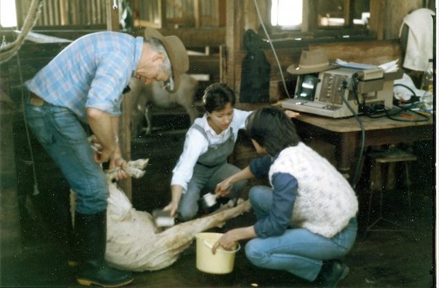
| 1992 | Sponsored research undertaken for Meat Research Corporation |
| 1996 | Colour Doppler imaging system licensed to Philips for use in their commercial equipment |
| 1997 | Staff split between CSIRO Marsfield and Lindfield |
| 2005 | Doppler flow measurement system licensed to Philips for use in their commercial equipment |
While the Ultrasonics Institute and its collaborators dominated the earliest years of ultrasound development in Australia, a number of pioneering Australian clinicians were quick to recognise the potential of the new modality and decided to explore it for themselves. Notable amongst these were*:
- Dr Ian McDonald (Melbourne, 1966)
- Dr John McCaffrey (Brisbane, 1966)
- Dr John Morrison (Brisbane, 1970)
- Dr Jim Roche (Sydney, 1971)
- Dr David Cooper (Brisbane, 1972)
- Dr Don Aickin (Melbourne, 1972)
- Dr John Stewart (Auckland, 1972)
- Drs Dugdale and Wong (Melbourne, 1973)
- Drs Cook, Furness and Verco (Adelaide, 1973)
- Dr Stan Reid (Perth, 1973)
- Drs Corrie, Taylor and Campbell (Hobart, 1973)
* Information from: Diagnostic Ultrasound, Peter Verco, Australasian Radiology 1981; 25th Anniversary Issue 115-123.
| 1965 | First demonstration of fetal spine, allowing identification of head from trunk and limbs and leading to determinations of fetal size and growth |
| 1966 | Demonstration of fetal intracranial echoes |
| 1968 | Demonstration of fetal orbits, heart, bladder, kidneys and scrotum |
| 1970 | Introduction of grey scale ultrasound, improving dramatically the demonstration of normal and abnormal structures in the fetus, placenta and mother |
| 1976 | Establishment of criteria for recognising placental ageing using grey scale ultrasound |
| 1976 | First discussion of fetal surgery |
| 1980 | Display of fetal brain development by grey scale ultrasound |
| 1980 | Monitoring of fetal umbilical venous blood flow in normal and complicated pregnancies |
| 1980 | Demonstration of fetal lung, liver and bowel maturation by grey scale ultrasound |
| 1993 | Development of real-time quasi-3D obstetric ultrasound |
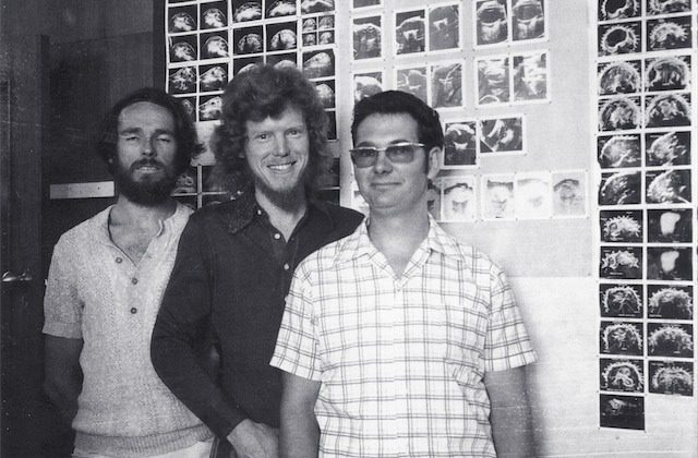
| 1974 | Grey scale ultrasound atlas of normal infant brain produced |
| 1975 | Assessment of hydrocephalus |
| 1978 | Abdominal grey scale ultrasound in children |
| 1980 | Production of tables and graphs of normal cerebral ventricular sizes in children |
| 1980 | Demonstration of detailed posterior cranial fossa anatomy in the neonate |


